New paper details the association between haemodynamic & electrophysiological brain signals
A new study conducted by Golia Shafiei, in the team of Bratislav Misic at The Neuro/McGill, in collaboration with our lab was just published in PLoS Biology. The study advances our comprehension of how functional MRI signals relate to electrophysiological fluctuations detected with MEG in brain networks.
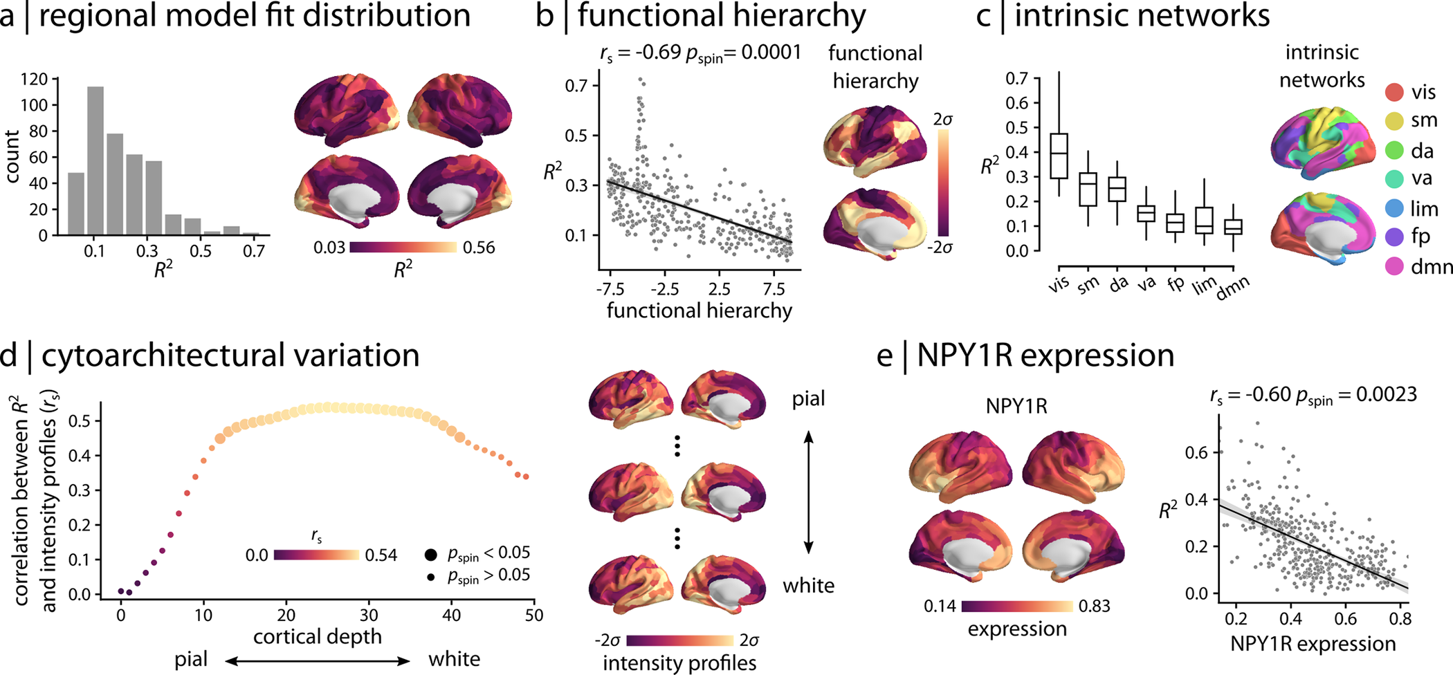
Whole-brain neural communication is typically estimated from statistical associations among electromagnetic or haemodynamic time-series. The relationship between functional network architectures recovered from these 2 types of neural activity remains unknown. Here, we map electromagnetic networks (measured using magnetoencephalography (MEG)) to haemodynamic networks (measured using functional magnetic resonance imaging (fMRI)). We find that the relationship between the 2 modalities is regionally heterogeneous and systematically follows the cortical hierarchy, with close correspondence in unimodal cortex and poor correspondence in transmodal cortex. Comparison with the BigBrain histological atlas reveals that electromagnetic–haemodynamic coupling is driven by laminar differentiation and neuron density, suggesting that the mapping between the 2 modalities can be explained by cytoarchitectural variation. Importantly, haemodynamic connectivity cannot be explained by electromagnetic activity in a single frequency band, but rather arises from the mixing of multiple neurophysiological rhythms. Correspondence between the two is largely driven by MEG functional connectivity at the beta (15 to 29 Hz) frequency band. Collectively, these findings demonstrate highly organized but only partly overlapping patterns of connectivity in MEG and fMRI functional networks, opening fundamentally new avenues for studying the relationship between cortical microarchitecture and multimodal connectivity patterns.